40 diagram of the eye without labels
Eye Diagram Unlabelled - schematron.org Best Human eye diagram unlabelled free vector download for commercial use in ai, eps, cdr, svg vector illustration graphic art design format. human eye. Ask A Biologistcoloring page | Web address:schematron.org coloring. Human Eye. Page 2. 5. 3. 2. 4. How to draw human eye in easy steps -10th -Physics - science - CBSE syllabus - NCERT class 10 Human Ear Diagram - Bodytomy The Structure of Human Ear. Helix: It is the prominent outer rim of the external ear. Antihelix: It is the cartilage curve that is situated parallel to the helix. Crus of the Helix: It is the landmark of the outer ear, situated right above the pointy protrusion known as the tragus. Auditory Ossicles: The three small bones in the middle ear ...
File:Schematic diagram of the human eye en.svg - Wikimedia Diagram of the human eye in English. It shows the lower part of the right eye after a central and horizontal section. ... Full redraw: Group labels in accordance with the "Foundational Model Explorer." Added "Macula" and "Uvea" and removed "Zonular fibres." ... File:Diagram of human eye without labels.svg; File:Figure of diplopia perception ...

Diagram of the eye without labels
The Eyes (Human Anatomy): Diagram, Optic Nerve, Iris, Cornea ... - WebMD The front part (what you see in the mirror) includes: Iris: the colored part. Cornea: a clear dome over the iris. Pupil: the black circular opening in the iris that lets light in. Sclera: the ... Galvanic Cell: Definition, Diagram and Working - Science ABC 17.01.2022 · Galvanic Cell Diagram. Now, consider this apparatus, which represents a galvanic cell. The first beaker contains zinc sulfate (ZnSO 4) into which a strip of zinc is dipped, while the adjacent beaker contains copper sulfate (CuSO 4) into which a strip of copper is dipped. However, the two strips are connected by an external circuit, a conductor, which is connected to a bulb. … Eye Anatomy: 16 Parts of the Eye & Their Functions - Vision Center The lens of the eye (or crystalline lens) is the transparent lentil-shaped structure inside your eye. This is the natural lens. It is located behind the iris and to the front of the vitreous humor (vitreous body). The vitreous humor is a clear, colorless, gelatinous mass that fills the gap between the lens and the retina in the eye.
Diagram of the eye without labels. eyeball diagram to label; Eye Cross Section Labeled Diagram Stock Vector - Illustration of we have 8 Images about Eye Cross Section Labeled Diagram Stock Vector - Illustration of like Medical Stock Art, Anatomy of the Eye, Human Eye Diagram, How The Eye Work -15 Amazing Facts of Eye and also Human Eye Diagram, How The Eye Work -15 Amazing Facts of Eye. Read more: Human Heart (Anatomy): Diagram, Function, Chambers, Location … WebMD's Heart Anatomy Page provides a detailed image of the heart and provides information on heart conditions, tests, and treatments. Anatomy of the eye: Quizzes and diagrams | Kenhub Take a look at the diagram of the eyeball above. Here you can see all of the main structures in this area. Spend some time reviewing the name and location of each one, then try to label the eye yourself - without peeking! - using the eye diagram (blank) below. Unlabeled diagram of the eye Create a Briliant Process Flow Diagram with Canva There are lots of ways to use color in a process flow diagram. You could have all the arrows in one part of the process the same color to make it clear they relate to that process. For example, you could use colors like blue and green to represent a cooling process or red and yellow to represent something being heated. To recolor any element or text in your design, select it, then …
Analyzing Minard's Visualization Of Napoleon's 1812 March 08.06.2014 · And he is able to show the drastic loss in life from Napoleon’s decision in just a single corner of the diagram. The beginning of Napoleon’s march vs the end of his retreat. Equally important as what’s shown is what’s not shown — here’s an earlier example of a well-published data visualization: Source. This graphic was created by William Playfair, largely … Anatomy of the Eye | Johns Hopkins Medicine The back part of the eye's interior. Pupil. The opening in the middle of the iris through which light passes to the back of the eye. Retina. The light-sensitive nerve layer that lines the inside of the back of the eye. The retina senses light and creates impulses that are sent through the optic nerve to the brain. Sclera. Eye Diagram Teaching Resources | Teachers Pay Teachers Anatomy of the Eye Diagrams for Coloring/Labeling, with Reference and Summary by Homemade For Play 8 $1.95 PDF This printable contains 13 clear and simple cross sectional diagrams of the human eye. They photocopy well and are great for use as a labeling and coloring exercise for your students. PDF Parts of the Eye - National Eye Institute | National Eye Institute Eye Diagram Handout Author: National Eye Health Education Program of the National Eye Institute, National Institutes of Health Subject: Handout illustrating parts of the eye Keywords: parts of the eye, eye diagram, vitreous gel, iris, cornea, pupil, lens, optic nerve, macula, retina Created Date: 12/16/2011 12:39:09 PM
Label the Eye - The Biology Corner Label the Eye Shannan Muskopf December 30, 2019 This worksheet shows an image of the eye with structures numbered. Students practice labeling the eye or teachers can print this to use as an assessment. There are two versions on the google doc and pdf file, one where the word bank is included and another with no word bank for differentiation. Free Pie Chart Maker - Make a Pie Chart in Canva A pie chart, also known as a circle chart, is a circular diagram that resembles a pie. Each of the ‘slices’ represents a category of data that makes up the whole. Together, the pie represents 100 percent. The size of each ‘slice’ is relative to its portion of the whole. Pie charts can become overly complicated if there are too many categories of data being presented. A good pie chart ... Human eye - Wikipedia The human eye is a sensory organ, ... Schematic diagram of the human eye. It shows a horizontal section through the right eye. ... Right eye without labels ... Eye Diagram With Labels and detailed description - BYJUS A brief description of the eye along with a well-labelled diagram is given below for reference. Well-Labelled Diagram of Eye The anterior chamber of the eye is the space between the cornea and the iris and is filled with a lubricating fluid, aqueous humour. The vascular layer of the eye, known as the choroid contains the connective tissue.

12 Best Images of Label Volcano Parts Worksheet - Volcano Diagram ...
Consumer Updates | FDA The site is secure. The https:// ensures that you are connecting to the official website and that any information you provide is encrypted and transmitted securely.
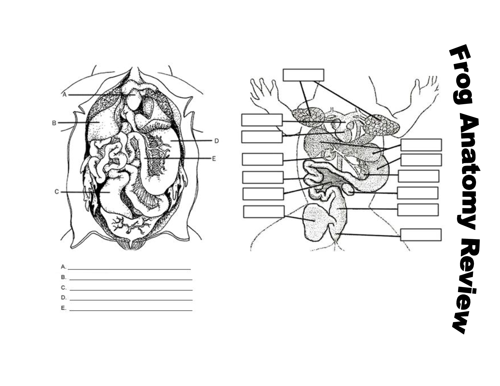
13 Best Images of Frog Anatomy Labeling Worksheet - Frog Dissection ...
The Eye Diagram: What is it and why is it used? The eye diagram is used primarily to look at digital signals for the purpose of recognizing the effects of distortion and finding its source. To demonstrate using a Tektronix MDO3104 oscilloscope, we connect the AFG output on the back panel to an analog input channel on the front panel and press AFG so a sine wave displays.
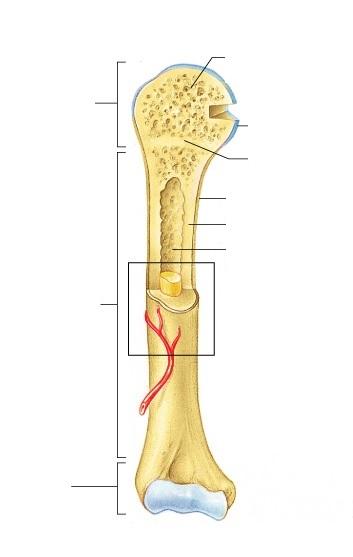
Exercise 9: Overview of the Skeleton: Classification and Structure of ...
Consumer Updates | FDA Science-based health and safety information you can trust.
Human Anatomy Review and Question Database
Your Eyes (for Kids) - Nemours KidsHealth It is a very important part of the eye, but you can hardly see it because it's made of clear tissue. Like clear glass, the cornea gives your eye a clear window to view the world through. Iris Is The Colorful Part. Behind the cornea are the iris, the pupil, and the anterior chamber. The iris (say: EYE-riss) is the colorful part of the eye. When ...
Post a Comment for "40 diagram of the eye without labels"