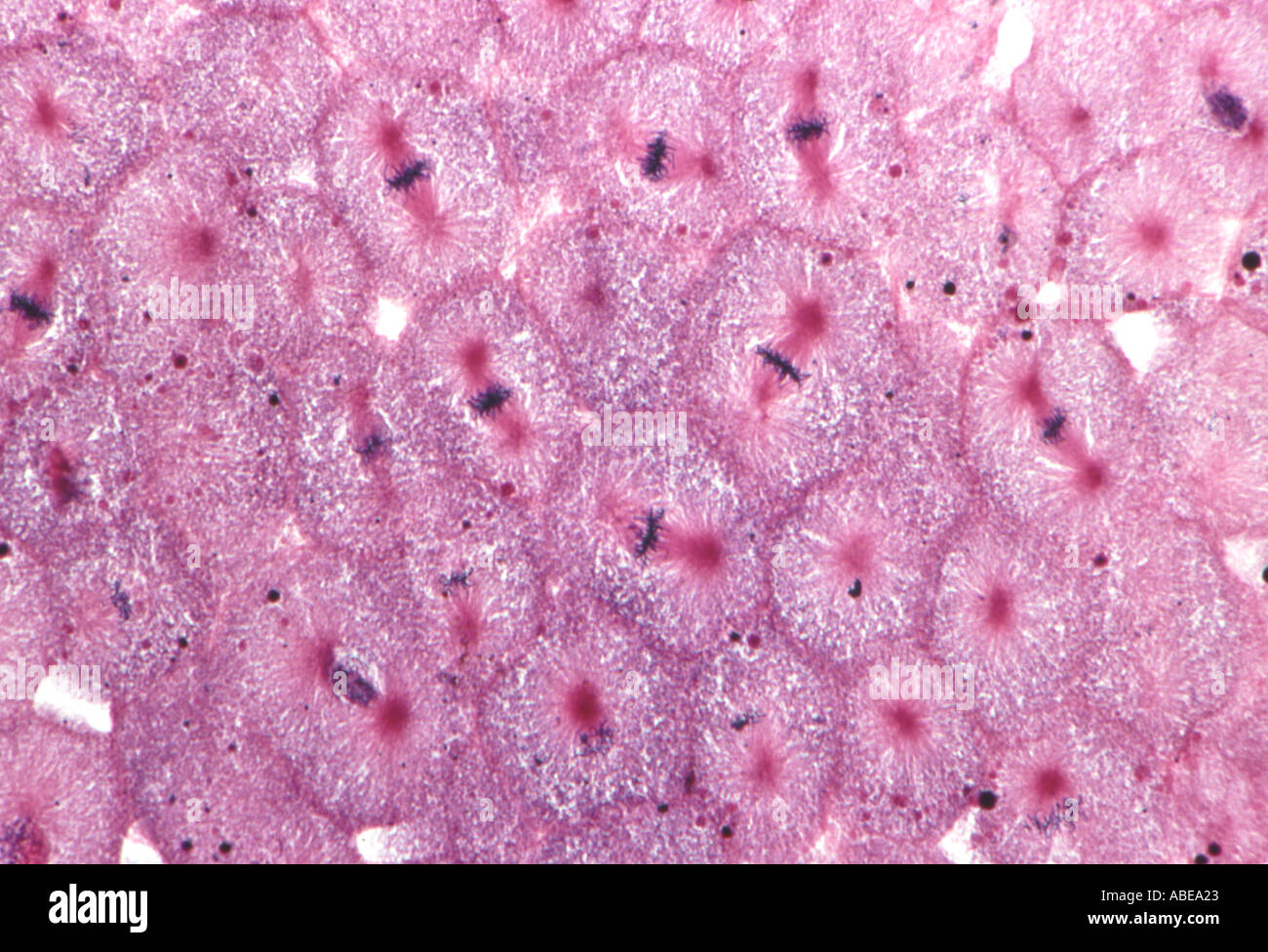
39 fish blastodisc mitosis
Histology Slides for Fish embryo cells; Animal; Whitefish blastodisc ... Histology Slides for Fish. Agnatha, chondrichthyes, and osteichthyesmitosis, coregonus (whitefish), blastodisc; mitotic stages are all clearly depicted. Manufacturer: Triarch Inc ZL6111. View more versions of this product. Catalog No. S11465. $17.00 / Each. Qty Check Availability. blastodisc | Encyclopedia.com blastodisc (germinal disc) A disc-shaped layer of cytoplasm that is formed at the animal pole by the cleavage of a large, yolky egg (e.g. that of a bird or reptile). Mitosis within the blastodisc gives rise to the embryo and its membranes. A Dictionary of Zoology
10 Mitosis and Meiosis | Laboratory Manual For SCI103 Biology I at ... Cut off a root tip and place it on a clean slide. Cut off 1mm to 2mm of the root tip and throw away the upper portion of the root. Cover the root tip with four drops of 1 N HCl and warm the slide over an alcohol burner flame for 1 minute. Do not boil. Blot off the excess HCl and cover the root tip with 0.5% aqueous toluidine blue.

Fish blastodisc mitosis
Mitosis in Real Cells - The Biology Corner Two specimens are commonly used by biologists to study mitosis: the blastula of a whitefish and the root tip of an onion. The whitefish embryo is a good place to look at mitosis because these cells are rapidly dividing as the fish embryo is growing. The onion root is also a good place because this is the area where the plant is growing. Fish & Onion Mitosis | Boreal Science Compare and contrast mitosisVWR offers slides for the varied purposes of your lab. Prepared and digital microscope slides for educational purposes are featured in an array of fields. General purpose microscope slides and cover glasses are offered as well as cavity, chamber, adhesion, and microarray slides for more specific research needs. Disposable and reusable options of varying thickness ... Mitosis Stages - Overview of the Stages of Mitosis - BYJUS The stages of mitosis comprise: Interphase Prophase Prometaphase Metaphase Anaphase Telophase Interphase Technically, the interphase is not a part of mitosis, however, it is still a crucial process as it leads up to the process of mitosis. Hence, the interphase refers to all the other stages of cell cycle other than mitosis. Prophase
Fish blastodisc mitosis. Fish Blastodisc, sec. 7 µm H&E Microscope Slide | Carolina.com Fish Blastodisc, sec. 7 µm H&E Microscope Slide | Carolina.com. My Account. Login or register now to maximize your savings and access profile information, order history, tracking, shopping lists, and more. Login Create an Account. Mitosis Blastodisc Prepared MIcroscope Slide ZL6-111 Mitosis Blastodisc Prepared Microscope Slide Mitosis. Agnatha, chondrichthyes, and osteichthyesmitosis, coregonus (whitefish), blastodisc; mitotic stages are all clearly depicted & are present in numerous quantities. Iron hematoxylin stain, section. A 10% discount applies if you order more than 10 of this item and 15% discount applies if ... Fish Blastodisc Mitosis sec_40X - PetroArc International Fish Blastodisc Mitosis sec_40X - PetroArc International. Solved LAB 10 Mitosis and Meiosis 10.2 Animal Cell Mitosis | Chegg.com 4. Name the two stages of mitosis when each chromosome is single-stranded (consists of one chromatid or monad). Anaphase Telophase + Cytokinesis Mitosis in Fish Blastodisc 3. Use the diagram of mitosis in an animal cell below to answer the following questions: Interphase Prophase Astor 5. Mitosis results in two identical cells.
Whitefish Mitosis - Green River College A whitefish blastula cell in anaphase During anaphase the mitotic spindle apparatus pulls the sister chromatids of each chromosome apart by attaching to each centromere and then pull the chromatids to each pole of the cell. Note that the telomeres of each chromosome point toward the cell's equator. Telophase Fish Blastodisc Mitosis - blastula stock photos and pictures getty ... Fish Blastodisc Mitosis - 14 images - anaphase animal mitosis 250x stock photo getty images, marine petroarc international, ppt whitefish blastula powerpoint presentation free download id 2373110, ascaris mitosis slide, What is the difference between onion and whitefish cells and the cells ... For mitosis experiments, onion root tips are used. The root tip cells are actively dividing meristematic cells and mitosis can be observed in them very easily. Cells in the testes are germ cells. And meiosis can be demonstrated only in germ cells; especially the stages of prophase I like crossing over and chiasmata can be easily seen in ... Fish Mitosis Slide | Ward's Science Fish Mitosis Slide: Ratings: Total Ratings: 0 Avg. Ratings: 0.0 out of 5. Sort all by: WARD470182-554. 470182-554EA 22.09 USD. 470182-554. Fish Mitosis Slide ... Nine typical sections of a blastodisc from several different eggs to show many mitotic figures at various stages of mitosis. ...
Fish Development - University of Calgary in Alberta Fish eggs are telolecithal. The large quantity of yolk restricts the mitotic apparatus and cleavage furrows to the blastodisc, a small, yolk-free zone at the animal pole. Consequently, the egg is not completely divided into individual blastomeres. Rather, they divide by discoidal cleavage. The embryo proper is formed only from the blastodisc. PDF 1/23/2004 Lab 3 Mitosis and Meiosis (pp 29-35 lab manual) 1. We will study mitosis and meiosis by looking at the stages of mitosis on prepared slides using the onion root tip, fish blastula, Ascaris ovary, and grasshopper testis. a. The onion root tip is fast growing and therefore have many cells in different stages of mitosis, but be advised that there are many cells not undergoing mitosis on these ... lab 5.pdf - 1 Fill-in the Table compare and contrast Mitosis vs Meiosis ... View Lab Report - lab 5.pdf from BIOL 160 at Green River College. 1. Fill-in the Table: compare and contrast Mitosis vs. Meiosis QUESTION MITOSIS MEIOSIS In what type of cell does each form of cell Fish Blastodisc Mitosis, 400X sec - PetroArc International Mitosis is a part of the cell cycle in which chromosomes in a cell nucleus are separated into two identical sets of chromosomes, and each set ends up in its own nucleus. In general, mitosis (division of the nucleus) is often accompanied or followed by cytokinesis, which divides the cytoplasm, organelles and cell membrane into two new cells containing roughly equal shares of these cellular components.
Whitefish Mitosis Whitefish Embryo High-Res Stock Photo - Getty Images
Fish, Blastodisc | VWR 470177-558EA 14.99 USD. 470177-558. Fish, Blastodisc. Slides Prepared Slides. Explore mitosis and genetic structures. Identifying features clearly distinguishable. Rigorous quality control standards. Demonstrating mitosis, asters, spindle fibers, and centrioles. New Product.

17b | Fish Blastodisc Mitosis with 10x lens | UAF Center for Distance ...
Fish, Blastodisc | Sargent Welch Explore mitosis and genetic structuresVWR offers slides for the varied purposes of your lab. Prepared and digital microscope slides for educational purposes are featured in an array of fields. General purpose microscope slides and cover glasses are offered as well as cavity, chamber, adhesion, and microarray slides for more specific research needs. Disposable and reusable options of varying ...

Cell Cycle and Mitosis, Laboratory Notes for BIO 1003
White Fish Blastula- Slide WSU_3_092 Prophase (A & C) is the stage of mitosis where the chromosomes contract linearly and thicken. The centriole divides and the two daughter centrioles migrate to opposite poles. Prophase comes from the greek word prophasis, which means to foreshadow. This is appropriate, because these changes are foreshadowing the division of the cell!

Mitosis Cell Division Chart | Carolina.com
Fish Mitosis 800.228.7117. All Categories All Categories Activity Sets & Kits

Fish Blastodisc Mitosis, 400X sec – PetroArc International
Cell Division - Mitosis - in Whitefish Blastula Cells Cell Division - Mitosis - in Whitefish Blastula Cells This cell is in the interphase stage of the cell cycle. Early in interphase the cell (A) reaches its full size and then starts preparing for its next division.
Mitosis in section of whitefish embryo Simultaneous division Stained ...
Cell Division - Mitosis - in Whitefish Blastula Cells Cell Division - Mitosis - in Whitefish Blastula Cells The arrow (A) points to a cell nearing the end of telophase. During telophase the spindle is disassembled and the nuclear envelope reforms around the divided sets of chromosomes. The appearance of a cleavage furrow (1) marks the onset of cytokinesis and the end of the process of mitosis.

whitefish mitosis | Mitosis, Anatomy and physiology, Physiology
Blastodisc - an overview | ScienceDirect Topics Blastodisc formation takes advantage of the pre-existing biased distribution of cytoplasmic and yolk components of the oocyte. After egg activation, this bias is enhanced by the animal pole-oriented streaming of ooplasm, a process that results in the separation of ooplasm from the yolk and lifting of the animal cortex to form a one-cell embryo.

Wall Chart - Mitosis - Biologyproducts.com
Whitefish embryo - The Biology Corner Whitefish Embryo [ Start Page | White Fish Page | Root Tip Page] Click on each image to enlarge. 4. For each image, sketch the cell that is highlighted and identify the stage the cell is in.
Mitosis Slide Whitefish Blastodisc Prepared Microscope Slide
Fish, Blastodisc | Boreal Science 470177-558. Fish, Blastodisc. Slides Prepared Slides. Explore mitosis and genetic structures. Identifying features clearly distinguishable. Rigorous quality control standards. Demonstrating mitosis, asters, spindle fibers, and centrioles. New Product.
Post a Comment for "39 fish blastodisc mitosis"